Stepping up the printing game: the first 3D heart from human tissue
When Gutenberg invented the printing press in 1450, he had no idea how far printing technology would have developed in the following centuries. Nowadays, any document can be easily and quickly printed in different colours with just a click. Imagining that one day we would be able to print a 3D heart with human tissue – this would not only astonish Gutenberg, but also sounds like a futuristic reality for some of us in 21st century. Yet, just last month, researchers at Tel Aviv Institute of Research 3D-printed the first heart using patients’ biological material1. So, let’s take a step back and understand how 3D printing works.
The process of 3D printing begins with the production of a CAD (computer-aided design) model. This is a 3D model used as blueprint to send specific instructions from the computer to the printer about the amount, location and type of material to use. By a procedure known as additive manufacturing, successive thin layers of material are precisely laid by a print head into the desired 3D shape. A variety of different materials can be used, including ceramics, polymers and metals to produce objects that vary in size, from jewellery to chairs or entire houses2.
3D printers are widely used in the medical sector. For example, the creation of 3D model organs allows surgeons to practice difficult operations in advance to dramatically reduce surgery time on patients. However, it is only recently that 3D printing technology has been applied to the field of regenerative medicine. Regenerative medicine aims to isolate and culture human cells in biomaterials, and engineer them to become functional tissues which can be used to replace damaged organs3.
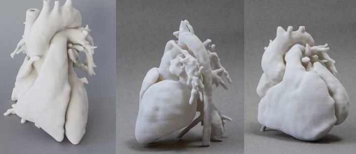
The leaders of the research at the University of Tel Aviv used this approach to 3D-print the first heart from patients’ own cells with the blood vessels, chambers, valves and cells like a normal human heart. In this medical breakthrough, a sample of the omentum (fatty tissue which protects the abdominal organs) was extracted from patients4. The cells were separated from the extracellular matrix, an aggregate of proteins and scaffold molecules which is arranged in an organised network between cells. Subsequently, the cellular material was re-programmed by inducing expression of specific genes and in doing so, it was transformed into stem cells with the ability to differentiate into various cell types. The extracellular matrix was processed to create a hydrogel. This is essentially a 3D structure made of polymers – large molecules made of many individual subunits. This can imitate natural tissues’ properties, and can therefore be used as scaffold structures to provide guidance in cell differentiation and organisation5.
Once incorporated in the hydrogel, the stem cells gave rise to specialised cardiac cells and endothelial cells (cells lining the internal aspect of blood vessels). These were subsequently used as ‘bio-ink’ for 3D printing. The result was the production of vascularised pieces of cardiac tissue (or cardiac ‘patches’), ultimately assembled in a heart.
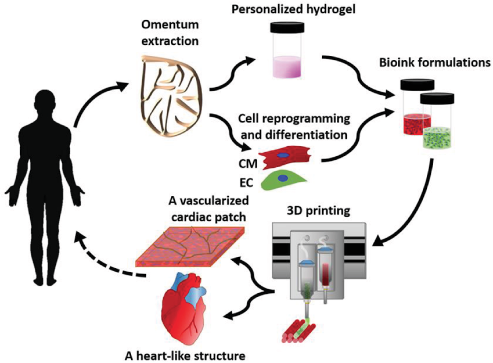
The procedure used by Tel Aviv researchers to 3D-print a heart from human tissue (EC = extracellular matrix, CM = cellular material)
Image credit: Dvir, T. et al., licensed under CC BY 4.0
The creation of a 3D-printed heart could have therapeutic potential for disorders affecting normal cardiac function, such as heart diseases. These conditions can often only be treated with an ‘allogenic’ transplant, in which a heart from a donor replaces the patient’s dysfunctional heart. However, the number of donors is scarce, and even when transplantation is possible, obstacles remain. For example, around 20% of the transplanted hearts are rejected by the patient’s immune system, which recognises the new heart as foreign, increasing risk of death.
These issues could be overcome with 3D printing technology, enabling the production of tissue from the patient’s own cells (known as ‘autologous’ tissue). This patient-specific tissue matches the immunological and anatomical features of the patient’s heart and thus allows a safer transplant process.
Despite having the same structural features as the human organ, the 3D-printed heart produced by Tel Aviv University researchers is only cherry-size and lacks essential functional properties to make it available for therapeutic purposes. However, this medical breakthrough sets the path for further developments in regenerative medicine. Perhaps in ten years, transplants involving 3D-printed organs will be one of many routine procedures in hospitals. Perhaps, in a decade’s time, the current standard transplants will be looked back upon as today we look back at Gutenberg’s primitive printing.
For now, researchers should focus on “teaching” the 3D-printed heart needs how to behave like a human heart. It will take some time before doctors will be able to use 3D-printed hearts for organ transplant – but as we know, technology can develop pretty fast!
Edited by Frankie Macpherson
Author
References
- The study was published in Advanced Science: https://onlinelibrary.wiley.com/doi/full/10.1002/advs.201900344
- A simple explanation of 3D printing: www.explainthatstuff.com/how-3d-printers-work.htm
- More on how to 3D print human tissue: https://phys.org/news/2018-11-scientists-patients-cells-materials-fully.html
- https://medicalxpress.com/news/2019-04-d-heart-human-tissue-vessels.html
- Hydrogels in regenerative medicine: www.ncbi.nlm.nih.gov/pmc/articles/PMC4494665/