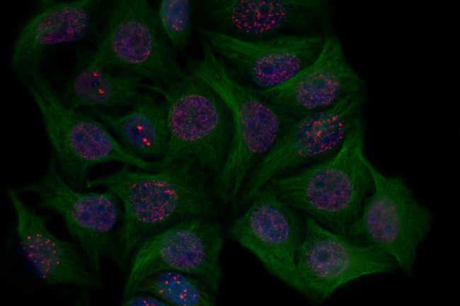
Approximately 8% of our genetic code can be traced back to ancient viral infections that occurred millions of years ago. Normally dormant in healthy cells, these genetic fragments contain instructions for viral proteins which are proteins that are normally only present on the surface of a virus particle and can be recognised by the body as being of foreign origin. These viral remnants were once part of infectious viruses long ago. The genes that code for them managed to endure and persist in the DNA of our ancestors, who survived viral infections and passed this genetic material down to us. These bits of our DNA typically remain unutilised. However, when a healthy cell undergoes a malignant transformation, its cellular machinery becomes chaotic, prompting cancerous cells to produce these viral envelope proteins. As a result, these long-forgotten viral remnants are awakened. Surprisingly, recent studies have unveiled the remarkable role of these once-considered ‘harmful’ proteins in triggering an immune response against cancer.
Viral envelope proteins have an important function: they are specific for each virus and oftentimes determine what the host cell of this germ will be. This unique set of proteins covers the genetic information of the virus and helps it attach to the host cell’s surface. A study revealed elevated levels of the Human Endogenous Retrovirus-7 (HERV-7) proteins in patients with lung adenocarcinoma, compared to non-malignant lungs and lung squamous cell carcinoma patients 1. This elevated expression of HERV-7 was found to be correlated with the formation of lung cancer. Notably, HERV-7 is recognised by the immune system as foreign, triggering the generation of an adaptive immune response and hence the production of antibodies, like immunoglobulin G (IgG), specifically compatible with HERV. This leads to a target destruction of the cell expressing the HERV protein 2.
Other studies have also highlighted a similar immune response to Endogenous Retrovirus-K (ERV-K), another viral envelope protein. The introduction of ERV-K-specific antibodies to the tumour site has shown potential in repressing the growth of cancerous cells. These findings support the role of ERV-K protein as a suitable target in immunotherapy. Positive patient responses have also been noted in other cancers; intriguing observations in renal cell cancer show activation of a region of chromosome six, coding for the virus Endogenous Retrovirus-E (ERV-E), which is associated with higher immunotherapy response 3.
The production of HERVs makes the tumour more susceptible to a type of immunotherapy, called Immune Checkpoint Blockade (ICB). Immune checkpoints are molecules on the surface of immune cells that play a crucial role in regulating the immune response by acting as ‘brakes’ that prevent immune cells from attacking healthy cells. In ICB therapy, immune checkpoint inhibitors prevent the binding of checkpoint proteins on T-cell immune cells, such as Programmed Cell Death Protein (PD-1), with the respective checkpoint proteins on tumour cells – PD-L1. This lifts the checkpoint ‘brake’ and allows the immune system to unleash an immune response against the cancerous cells and kill them. This recognition of tumours by the immune system is enhanced when HERV-derived, recognisable viral envelope proteins are present on their surface. Overall, HERV antigens play a pivotal role in promoting the immune response, attracting more T-cells to the tumour site.
Uninterrupted immune checkpoint protein binding between Programmed cell death protein (PD-1) and its counterpart (PD-L1) versus interrupted checkpoint by an immune checkpoint inhibitor. Image by Veneta Salyahetdinova created in Biorender.
HERV-based immunotherapy holds great potential in the fight against cancer. However, there are certain limitations of the method. For instance, one may expect some off-target effects, since therapies which target HERVs could affect healthy cells as well. Another thing to consider is the possibility of developing immune resistance to the HERV class antigens. HERVs present a unique challenge known as ‘viral mimicry’. In essence, the HERV protein may resemble other types of antigens, which could lead to a ‘false alarm’’ among immune cells and the subsequent activation of an unwanted autoimmune response.
What precisely activates the dormant expression of these long-forgotten viruses? Scientists propose that the upregulation of viral genes results in an overexpression of proteins derived from HERVs, leading to an increase in HERV-specific antibodies. Moreover, in cancer cells, reactivation of HERV elements may be due to abnormalities not in the genetic material itself, but in the way it was expressed; modifications in the so called epigenetic factors. Additionally, cellular stress and inflammation are believed to contribute to the synthesis of viral envelopes within cancer cells.
This research suggests that viruses can be surprising allies in our fight against cancer, challenging the long-standing belief that they are solely our enemies. Further investigation into the specific viral genes reactivated in different types of cancers is essential. The presence of viral envelope proteins appears to serve as a promising biomarker, an indicator of pathogenic processes, for certain cancer types, which opens new possibilities for diagnosis and targeted therapies.
Edited by Lydia Melissourgou-Syka
Copy-edited by Rachel Shannon

