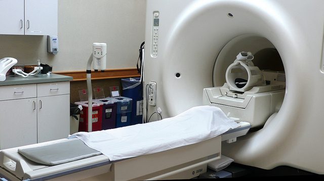
Focused ultrasound or magnetic resonance-guided high intensity focused ultrasound surgery (MRgFUS) technology is a non-invasive technique developed to target internal body tissues. In the 1950s, Francis and William Fry demonstrated that high intensity focused ultrasound could be used to target deep tissue in the brain of patients with Parkinsonism disorders 1. Since then, the technology has advanced significantly, and it can now be thought of as an alternative to incision-based surgery or radiation therapy.
Magnetic resonance imaging (MRI) is used to scan the area affected and detect the problematic tissue. Focused ultrasound energy is then used to heat the diseased or damaged tissue to destroy it by means of ablation. MRI allows the physician performing the surgery to control and monitor the entire procedure whilst it is being carried out.
Prior to surgery beginning, a transducer is attached to the affected area of the patient’s body. Transducers are electronic devices that can change one form of energy into another. The patient is then placed in a MRI scanner and a MRI scan performed. From this scan, the tissue which requires treatment can be identified. The target area can be anywhere in the range of 1 x 1.5 mm to 10 x 16 mm in size. The damaged or diseased tissue is then destroyed via thermal ablation. At low energy, the focused ultrasound wave does not damage the tissue. It can however, elevate the temperature of the tissue. The physician can use this change in temperature to ensure that the correct tissue has been selected before the procedure begins. The temperature of the tissue is raised to between 55 and 60 degrees Celsius for a few seconds, which causes the proteins and cells of the tissue to denature. The surrounding tissue is unaffected by the procedure.
Whilst not a widely known or used technique, focused ultrasound has been approved for use in the treatment of uterine fibroids in women since 2004. Research is ongoing into the application of focused ultrasound technologies for the treatment of neurological conditions, such as Parkinson’s disease, essential tremors, cancerous tumours and neuropathic pain. It is believed that the side effects often encountered when treating these conditions could be avoided when using non-invasive surgical techniques such as MRgFUS. For example, using MRgFUS to target prostate tumours would allow for the removal of the tumour without removing the prostate gland, as per the current procedure. As such, erectile dysfunction and incontinency issues associated with this surgery would be less likely to occur if a non-invasive alternative was employed. Focused ultrasound surgery also has reduced psychological effect on the patient, and has the added benefits of decreased recovery time and no scarring. It does however, have the disadvantage that complete removal of the tumour cannot be verified.
While the future of these applications is unknown, it is easy to imagine that focused ultrasound technology is an up and coming technology in the world of non-invasive surgery.
Edited by Debbie Nicol
References
- F.J. Fry, Am J Phys Med, Precision high intensity focusing ultrasonic machines for surgery

